Published On September 22, 2009
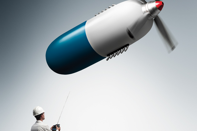
TAKING MEDICINE NEVER USED TO BE ROCKET SCIENCE. You swallowed a pill and it dissolved in the stomach, entered the small intestine and was absorbed into the blood. From there the drug dispersed throughout the body, delivering at least a little where it was needed. But many drugs are poorly soluble or are eliminated by the liver very quickly, so they have to be taken in high, potentially toxic concentrations to be effective. What’s more, how much of a drug gets absorbed depends on its physical and chemical properties, and some medicines may have trouble reaching their destination. For example, compounds that are fat soluble cross fat-containing cellular membranes easily but won’t dissolve in water. And biologic drugs—immune-system-stimulating marvels engineered from living organisms—tend to be composed of large molecules that can’t pass through the membrane of the small intestine and must be injected rather than swallowed. Getting drugs into the brain is even more daunting because cells lining the blood vessels there are packed so tightly they create a nearly impenetrable defense against particles trying to reach brain tissue.
As researchers and pharmaceutical manufacturers have devised ever more ingenious drug formulations, delivery issues have mushroomed. “The problems we face aren’t so much coming up with better drugs but making sure they go to the right place,” says Mauro Ferrari, chairman of the department of nanomedicine and biomedical engineering at the University of Texas Health Science Center in Houston. Ferrari, who trained as a mathematician and engineer, is one of a host of scientists from new disciplines—physics, engineering, robotics—confronting those conundrums. Working with nontraditional materials and technologies, these researchers are finding their way past the body’s intricate defenses to get more of today’s miracle medicines where they need to go.
Frequently that’s a life-or-death issue. The cancer drug Taxol (paclitaxel), for example, is poorly soluble unless mixed with a solvent that can cause life-threatening allergic reactions, requiring that patients be pretreated with steroids and antihistamines. Even when the best of anticancer drugs are injected, often only one molecule out of 100,000 hits its mark. That means many drugs have to be given frequently and at high doses, and that tends to cause side effects and toxicity.
Ferrari focuses on building nanosize drug carriers that burrow into a tumor before releasing their cargo of drug particles. Other scientists working in the nanorealm are covering drug particles with silicon wires that stick to slippery cells normally bathed in mucus, such as those in the nose and intestines. Another technology being tested is coating nanoparticles with a gel that explodes, sending the particles through the body at nearly 800 times normal speed to force them through thick tissue.
Older technologies, too, are being called to new purposes. Ultrasound has shown promise in lab animals for opening a temporary passage to brain tissue never before accessible to drugs. Microprocessors are being embedded in implantable devices that can be programmed to release precise doses of a drug. And a “pill” packed with sensors and a wireless transceiver can release medicine at just the right moment during the pill’s passage through the intestines. All these systems, though still experimental and probably years away from Food and Drug Administration approval, are part of a wave of drug delivery that may change how medicines find the body’s sweet spots.
NANOMEDICINE, USING MATERIALS THE SIZE OF MOLECULES, holds the promise of delivering drug particles (nanoparticles) so small they can avoid getting caught in the liver’s filter or can flow into tumors’ leaky, irregularly shaped blood vessels, which can trap larger particles. But even on this microscopic scale, shape matters, affecting the ability of nanoparticles to flow through capillaries and adhere to blood vessel walls. “Sphere-shaped particles have the lowest probability of landing in a tumor—and nearly all the particles we use are spheres,” Ferrari says. “So drug design becomes a hemodynamic engineering problem to create disklike, cylindrical or hemispherical particles, which fare hundreds of times better.”
The first generation of nanocarriers, developed in the mid-1990s, were liposomes, tiny spheres that could be filled with anticancer drugs. Some injected liposomes end up in the irregular blood vessels that spring up to nourish tumors, and the longer the liposomes remain in the blood, the more likely it is that some will get trapped in those defective capillaries. A portion of the liposomes do make it through the porous blood vessels and reach cancer cells in the tissue, where they are broken down by the cell’s metabolism and unleash their chemotherapeutic payload. This approach delivers as much as 50% more of the drug than would arrive if it were injected without the liposomes, but the window of opportunity is small. Administering a high concentration of a drug toxic to cancer cells may also degrade the blood vessels, causing them to collapse. Moreover, many cancer treatments now include anti-angiogenic drugs to kill or normalize the blood vessels feeding the tumor, cutting off liposomes’ easy access to the cells.
To improve the performance of liposomes and other nanoparticles, researchers have tried covering their surfaces with such agents as monoclonal antibodies, lab-produced substances designed to zero in on cancer cells and bind to their receptors. However, that approach has met with only modest success, in part because it’s difficult to get a sufficiently high concentration of a drug past the body’s defenses against foreign substances. Even nanoparticles may be stopped short by scavenging white blood cells, called phagocytes, that divert drug particles to the spleen and liver for disposal.
Still, Ferrari thinks he has a solution, a nanocarrier just one-hundredth the thickness of a human hair. “The nanocarrier has to avoid being attacked by the body’s immune system,” he says. “It has to chew through seemingly impenetrable cell membranes, survive a very acidic environment in the cell and overcome a tumor’s internal pressure, which tends to push drugs away from it. To make it into the treasure room—the cell’s nucleus—the nanocarrier needs to open all 10 doors. So you need a multistage nanocarrier that sequentially delivers different drugs performing different functions.”
Injected into the bloodstream, the first stage of Ferrari’s nanocarrier would bind to the inner wall of a blood vessel near the cancer cells, assisted by an antibody coating that seeks out the abnormal vessels around a tumor. As the first stage degrades, it releases the second—nanoparticles that burrow through the vessel walls and chew through the membrane on which the blood vessel cells sit—to reach the diseased cells.
Inside the tumor, the third stage is released, dispensing either medication to kill the cancer cells or a contrast agent that facilitates real-time imaging of the cancer, or both. To avoid phagocytes, Ferrari is experimenting with changing the size and charge of the nanoparticles so they won’t be mistaken for dying or dead red blood cells, which the phagocytes devour.
If Ferrari’s nanocarrier works for cancer, it might also be used to treat or diagnose other conditions, such as hemorrhages or heart disease, that create changes in blood vessels. “A nanocarrier with diagnostic capabilities could highlight the formation of coronary artery plaque, for example,” says Ferrari, who thinks nanotechnology can make any drug better. “Ten years down the road, I would be surprised to find a drug that’s not delivered through a nanosystem.”

SOME DRUG REGIMENS ARE SO ONEROUS or unpleasant that patients refuse to comply with them. That common problem undercuts the value of many therapies, says Michael Cima, professor of engineering in the department of materials science and engineering at the Massachusetts Institute of Technology, who builds implantable devices designed to make drug administration more bearable. For example, patients receiving chemotherapy for bladder cancer or lidocaine for the painful disorder interstitial cystitis must be catheterized so their bladder can be filled with a highly concentrated drug solution. “Then they have to hold the solution in for as long as possible,” says Cima. “For interstitial cystitis, the procedure is repeated every other day for two to six weeks.”
To provide an alternative, Cima has created a device that continuously forces a lidocaine solution out of a tiny hole in a silicone tube, delivering a constant, more therapeutic dose of the drug than is possible when a patient holds a stronger solution in her bladder for 30 minutes or so. The one-millimeter tube, inserted into the urethra, flares open so it won’t be carried out during urination. “The tubing is thin and permeable enough that urine can leak through it, causing the lidocaine, in the form of a salt, to dissolve,” Cima says. “Then the high salt concentration inside the tube creates an osmotic pressure that forces the solution out.”
Cima is also working on more complex, electronic devices, including one about the size of a blueberry that can be implanted under the skin. It provides controlled-release parathyroid hormone therapy for osteoporosis that would otherwise have to be injected daily. The drug is packed into reservoirs covered by very thin membranes of gold, which melt at comparatively low temperatures. An embedded microprocessor programs a pacemaker battery to deliver electric pulses to melt the membranes and release the drug, one reservoir at a time. The device, which is still being tested, can be programmed before it’s implanted or wirelessly controlled afterward.
Not all of Cima’s delivery devices release drugs slowly; some are specifically designed to provide rapid infusion after being activated by sensors that detect certain symptoms, such as the falling blood pressure and heart rate of a wounded soldier. People at risk for life-threatening events, such as severe allergic reactions, epileptic seizures or cardiac arrest, could have such a device implanted as a preventive measure. A model Cima has tested on rabbits has a titanium bottom layer lined with microresistors that quickly heat a Pyrex reservoir filled with a liquid drug, such as vasopressin, which restores blood pressure when a person is in shock. Vapor bubbles that form in the drug generate pressure that ruptures the membrane sealing the capsule, causing the drug to jet out into the body.
Whereas Cima’s devices must be implanted, other delivery systems can simply be swallowed. Inspired by capsule endoscopy—a camera-in-a-pill that photographs the small intestine, which can’t be readily imaged from outside the body—Dutch gastroenterologist Peter van der Schaar, working with biomedical device maker Philips, developed the intelligent pill, or iPill, to deliver medication to an inflamed or diseased portion of the intestines. Many gastrointestinal conditions, such as Crohn’s disease and colitis, are treated with steroids, which have adverse effects when they’re dispersed throughout the body. The iPill can deliver steroids or other drugs only where they’re needed.
The iPill contains a microprocessor, a battery, a fluid pump and a wireless radiofrequency transceiver. Less than an hour after being swallowed, the device leaves the highly acidic stomach and enters the alkaline small intestine. Registering the jump in pH levels, the microprocessor begins a countdown: During the next six hours, it triggers the pump to release medication when the elapsed time indicates that the capsule has reached a targeted region. The microprocessor can also be programmed to dispense the drug in multiple bursts every few minutes. If more precision is needed, physicians can watch the iPill’s transit with imaging technology (a CT scan yields an image accurate enough to show the iPill’s location) and send a radio signal to release the drug when the capsule passes a particular spot.
Jeff Shimizu, a senior scientist at Philips Research North America in Briarcliff Manor, N.Y., where the iPill is being further developed, first tested the device in a saltwater aquarium to make sure its radio signals would work in the intestines’ saline environment. Now he’s trying to reduce the size of the cylinder, which is about as big as a Magic Marker top. “It has been a real challenge to build a capsule small enough to swallow but with a large enough drug reservoir and the necessary sensors,” says Shimizu. “People look at it and say, ‘That’s a big pill.’”
BEING ABLE TO DELIVER DRUGS TO the usually inaccessible intestines is a major advance, but reaching the brain would be tremendous. Cells lining blood vessels elsewhere in the body normally have gaps that allow some drug molecules to pass through. But in the brain’s blood vessels, the cells adhere with extremely tight junctions, erecting the blood-brain barrier to keep out foreign substances. “Ninety-eight percent of traditional small-molecule drugs won’t pass through the blood-brain barrier, and none of the large-molecule drugs, such as monoclonal antibodies, make it through,” says Nathan McDannold, research director of the focused ultrasound laboratory at Brigham and Women’s Hospital in Boston.
McDannold and his mentor Kullervo Hynynen, now at Sunnybrook Health Sciences Centre in Toronto, had worked on a successful clinical trial using focused ultrasound to shrink uterine fibroids. They were intrigued by experiments done in the 1960s that used the same method to destroy tissues deep inside an animal’s brain. A contrast agent used in the earlier tests had unexpectedly spread from the blood to brain tissues. But when they tried to re-create the experiments in rabbits, they found that a focused ultrasound wave directed at blood vessels in the animals’ brains sometimes caused hemorrhaging and trauma. “The very intense ultrasound field created microbubbles from small gas nuclei in the blood,” explains McDannold. “The bubbles kept expanding until they burst, producing a violent shock wave that damaged brain tissue.”
So McDannold and Hynynen tried injecting the rabbits with an ultrasound contrast agent with preformed microbubbles that stay in the blood vessels. When they applied a low-intensity ultrasound beam, the results were instant and remarkable. “Using electron microscopy, we saw the contrast agent leaking through gaps that had opened between the cells,” McDannold says. Somehow the oscillating bubbles caused the gaps between cells in the blood-brain barrier to widen for about three hours, and there was no evidence of tissue damage.
The ability to open the blood-brain barrier temporarily in a small area could enable doctors to administer a high-concentration drug without fearing it might affect the rest of the brain. (The drug wouldn’t spread because that would require a reverse trip through the blood-brain barrier and into the bloodstream.) A student in his lab used this approach to give a single chemotherapy treatment to rats with brain tumors and found a modest increase in survival. Multiple treatments in people might be more effective, McDannold says.
The first focused-ultrasound drug treatment in humans will probably also be for a brain tumor. “Though there might be some mild unwanted effects from the ultrasound, such as slight vascular damage, that could be acceptable,” McDannold says, if it permits therapy for otherwise untreatable cancer. He speculates that the procedure might also be used to treat epilepsy, by putting a neurotoxin in a localized area to destroy the locus of the seizures in the brain. And his laboratory has done animal experiments in which the blood-brain barrier was opened to allow entry of an agent that detects and marks the amyloid plaques of Alzheimer’s disease. Current clinical trials are using focused ultrasound to destroy prostate, bone, breast and liver tumors, but McDannold thinks the technology’s ability to provide access to brain tissue could have a more profound impact. “There are so many drugs to treat neurological disorders that you can give using this technique,” he says.
That’s the point of alternative drug delivery systems—to deliver medications where they can be most effective. “We’re beyond the concept of creating new drug carriers,” says Robert D. Arnold, assistant professor of pharmaceutical and biomedical sciences at the University of Georgia College of Pharmacy. “The goal now is to develop drug carriers that will target the right site, deliver the payload at exactly the time the cell is most receptive to the drug and contain a probe that will allow us to see whether the drug has reached its target. When we can do all that, we’ll truly achieve optimal therapeutics.”
Dossier
“The Next Generation of Drug-Delivery Microdevices,” by N.M. Elman et al., Clinical Pharmacology & Therapeutics, May 2009. This overview visually illustrates the ingenious technologies being used to treat acute and chronic diseases as well as trauma.
“Progress and Problems in the Application of Focused Ultrasound for Blood-Brain Barrier Disruption,” by Natalia Vykhodtseva, Nathan McDannold and Kullervo Hynynen,Ultrasonics, April 14, 2008. Having built upon a 50-year-old finding that an ultrasound beam can disrupt the blood-brain barrier, the authors describe recent research showing that drugs can be targeted at selected areas.
“Five Big Ideas for Nanotechnology,” by Jon Evans,Nature Medicine, April 2009. Evans examines five scientists’ concepts for drug-containing nanoparticles, from particles coated with nanowires that stick to cells like burrs to particles that release different drugs when exposed to different wavelengths of light.
Stay on the frontiers of medicine