Published On May 3, 2008
JENNIFER AND DOUGLAS ROSE KNEW they were expecting twins, but a routine ultrasound at 20 weeks of gestation showed the girls, who shared a placenta, weren’t developing properly. Some blood vessels that joined the placenta were abnormal, resulting in a disproportionate circulation of blood to the fetuses. One was surrounded by an overly large amniotic sac, and the other’s sac was too small. What’s worse, the fact that the condition, known as twin-twin transfusion syndrome, or TTTS, was visible so early in the pregnancy meant it was especially acute. Untreated, one baby would receive too much blood, become hypertensive, urinate excessively and produce an overabundance of amniotic fluid. The other twin would suffer from low blood pressure, a deficit of oxygenated blood, kidneys that don’t produce urine and stunted growth. Both babies would likely die of heart failure.
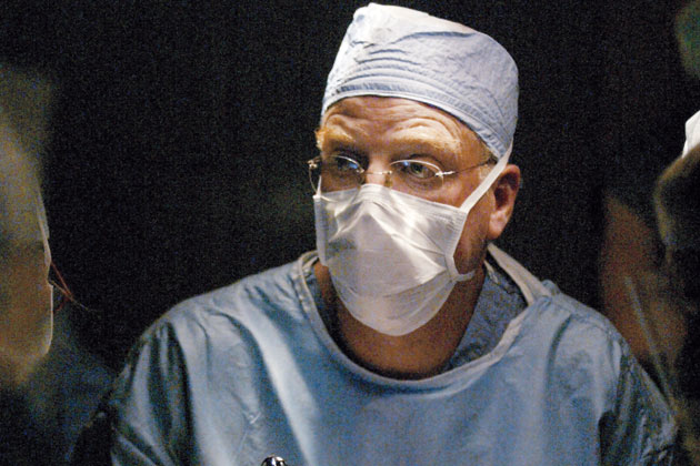
The conventional therapy for TTTS, amnioreduction, uses a needle to remove as much as three liters of amniotic fluid from around the twin with too much blood and in some cases may prevent the condition from progressing, though it can’t reverse its effects. But that approach would have had little effect in this severe case, and within days of her ultrasound on Sept. 25, 2006, Jennifer Rose, 38, was at the Children’s Hospital of Philadelphia to be evaluated for an alternative treatment. Mark Johnson, the hospital’s director of obstetrics for the Center for Fetal Diagnosis and Treatment, performed a procedure, selective laser ablation, on the abnormal vessels connecting the twins. “A huge system of blood vessels had expanded over the surface and within the placenta, much like the roots of a tree,” says Johnson. His plan was to insert a two-millimeter fetoscope equipped with a laser into Rose’s abdomen and map every vessel from its origin in the umbilical cord to the point at which it joined the placenta, then use the laser to close off any vessel that improperly connected the twins.
Rose decided to have the surgery, despite considerable risks to the fetuses. “We were told there was a 40% chance both babies would survive, and a 60% chance that one would,” she says. The anterior position of her placenta further complicated things, partially blocking Johnson’s view. But he managed to close off the problem blood vessels, and after a two-day hospital stay and 10 days of bed rest in Philadelphia, Rose went home to Waterbury, Conn., to await delivery. During further bed rest, she couldn’t pick up her infant twins, who had been born less than a year earlier. Frequent ultrasounds showed the developing fetuses and their amniotic sacs to be roughly the same size until 35 weeks into Rose’s pregnancy—just two weeks short of full term—when one baby suddenly gained weight and began producing too much amniotic fluid. Rose’s obstetrician induced labor a week later, and Rose vaginally delivered two healthy girls, 6 pounds 9 ounces, and 5 pounds 8 ounces, whom the parents named Faith and Grace. “Before this surgery existed, these babies would not have been born,” says Rose.
Of the approximately 100 fetal procedures done annually at the Children’s Hospital of Philadelphia, half are for TTTS, which has become increasingly prevalent because of the rising use of fertility drugs. The surgery is one of a half-dozen or so interventions that have been tried in any numbers since the 1980s, and it’s among the few that have proved clearly superior to the alternative of waiting until after birth and then operating only if necessary. Yet, while the idea of detecting and fixing problems before birth has long been appealing, and experiments have been under way for decades, just a handful of randomized clinical trials have even attempted to compare the two approaches. That’s partly because parents often refuse to participate for fear their fetus will be assigned to the control arm of a study and thus excluded from an intervention that could save or prolong the fetus’s life. And it’s not always clear which fetuses will benefit from in utero surgery, an uncertainty that may expose babies and their mothers to risks without potential rewards.
Still, while slow progress has discouraged some physicians, many are pushing ahead with their work on fetal interventions for congenital diaphragmatic hernia (CDH), hypoplastic left heart syndrome (HLHS) and myelomeningocele (spina bifida). The last is far more common than the other conditions, and surgery to correct it was the most frequent open fetal procedure, with some 200 operations performed before the work was suspended at all but three hospitals that are now participating in a major randomized trial. It has also been the most hyped, touted by grateful testimonials from parents on the Internet sites of fetal surgery centers across the country. Demand had become so great that fetal experts worried the surgery would become routine before any benefits were found.
That’s why everyone involved in fetal surgery is anxiously awaiting results of the spina bifida trial, which is expected to take several years. But by then, doctors may be attempting to fix fetal defects in entirely new ways: stem cell transplants and gene therapy. By then as well, some of today’s fetal procedures may have been abandoned amid ongoing questions of whether they are really worthwhile. Already on its way out (at least in the United States) is one of the earliest interventions: CDH repair. “Treating after birth, we get nearly 100% survival in babies with mild CDH and 70% in those with severe cases,” says Russell Jennings, director of the Advanced Fetal Care Center at Children’s Hospital Boston and a research fellow on the first team to do the procedure in utero. “So what’s the point?”
IN 1969, AT THE MASSACHUSETTS GENERAL HOSPITAL, after helping fix a hernia in a newborn’s diaphragm and then watching the baby die following an all-night vigil, surgical intern Michael Harrison had a big idea: repairing CDH in utero. The hole in the diaphragm wasn’t the real problem; rather, it was the abdominal organs that squeezed into the chest cavity, leaving no room for lungs to develop, so surgery on the fetus might allow the lungs to grow normally. At the time, the only fetal procedures that had been attempted were blood transfusions for Rh disease. Yet the thought that a fetus could be a patient never left Harrison, and in 1978 he joined the staff of the University of California, San Francisco, attracted by the research of pediatric cardiologists who were using a fetal sheep model to study the physiology of a newborn’s congenital heart defects. “But when I said, ‘Why don’t we think about fixing the heart in utero?’ they about fell off their chairs,” Harrison recalls.
Harrison pressed ahead, and in 1984, after 2,000 operations on fetal sheep and 500 on fetal monkeys, he tried a CDH repair on a human fetus. The surgery was radical—“I would just cut and sew in a big, terrible, open operation,” Harrison says—and no fetuses survived until 1989. But once he’d had a few successes, Harrison applied for funding from the National Institutes of Health for a randomized controlled trial to compare fetal surgical repair of CDH with postnatal surgery. The results, based on interventions involving just 11 fetuses and newborns, were “disappointing but interesting,” as Harrison reported in 1997. Those treated in utero had a 75% survival rate, compared with 86% for repair of CDH after birth. In such a small study, those outcomes were statistically indistinguishable.
With such questionable benefits, Harrison and other surgeons stopped doing that surgery but continued their research. As minimally invasive surgical techniques were developed for adults, fetal surgeons miniaturized endoscopes and other instruments, adapting them for their own use. Improvements in ultrasound made it easier to diagnose fetal problems and visualize the surgery, and doctors discovered that deeper anesthesia for the mother relaxed the uterus during surgery and helped prevent preterm labor. Surgery became less invasive and safer for both mother and child.
Still, CDH interventions in the womb have yet to prove as beneficial as operating on a newborn. From 1999 through 2001, a second NIH randomized trial involved 24 fetuses and newborns. But once again, the results favored the neonatal approach, which had a 77% survival rate compared with 73% for the fetal intervention. Further tipping the scales was that fetuses undergoing the procedure were delivered at 31 weeks, six weeks earlier than babies who were operated on after birth. As experiments continue, particularly in Europe, U.S. surgeons have largely abandoned fetal CDH repair.
Results have been much better for twin-twin transfusion syndrome. Compared with the conventional treatment of amnioreduction, selective laser ablation is less likely to prompt preterm delivery; fetuses are typically born at 33 to 34 weeks vs. 28 to 31 weeks with amnioreduction. Those extra weeks in utero reduce the risk of neurologic and cognitive problems—including mental retardation, cerebral palsy and muscle weakness disorders—that afflict as many as a third of babies who have amnioreduction. “Those risks fall to 7% or 8% with the laser procedure,” Johnson says. “That’s a significant difference.”
In the early 1990s, when surgeons first performed TTTS treatments, their tools were large and clumsy, but European researchers demonstrated that laser treatment of TTTS in the womb is significantly better than amnioreduction. The results reported in a 2004 European trial were so striking that a clinical trial under way in the United States was abruptly halted. Nearly 48% of the babies who had the fetoscopic laser procedure were alive and neurologically sound at six months compared with only 26% of those who had had amnioreduction.
“It was the first randomized controlled trial that showed any benefit for a fetal intervention, which was fantastic,” says Nick Fisk, a fetal-medicine specialist and director of the University of Queensland Centre for Clinical Research in Brisbane, Australia. “And initially, laser ablation appeared also to significantly reduce neurological injury in neonates. But there was no long-term reduction of cerebral palsy rates.”
Moreover, says Fisk, the procedure is still far from perfect. Trial results suggest that two-thirds of the time, laser ablation leads to brain damage or death for at least one twin. Meanwhile, 70% or more of twins with the mildest form of TTTS do better with no intervention or a single amnioreduction, and predicting which fetuses will progress to a more severe stage after 25 weeks (the cutoff for laser ablation because of the risk of rupturing vessels) is extremely difficult. “So you’re left with the question of how to treat early-stage disease,” Fisk says.
One answer may come from researchers at Children’s in Philadelphia who have just developed a technique involving echocardiograms and Doppler ultrasound measurements that can evaluate the severity of TTTS and the likelihood it will progress. “With a new, 20-point scoring system, we can observe a characteristic series of changes that occur in the vessels and the heart that tell us this is TTTS and not something else,” says Johnson. “It lets us know when to intervene and how to tailor treatment more effectively.”
Knowing whether to intervene in utero to repair hypoplastic left heart syndrome, a grossly underdeveloped and nonfunctioning left ventricle and aortic valve, is murkier yet. The rare heart defect is detected during routine ultrasound in fewer than half of the fetuses that have it. And some cases of HLHS are better dealt with after birth, if at all. “HLHS surgery is sensational, but no one knows which babies need it and which don’t,” says pediatric surgeon Alan Flake, a fetal-surgery pioneer and director of the Children’s Center for Fetal Research at the Children’s Hospital of Philadelphia. “I’m not knocking it—I’ve been in this field a long time and think it’s a good thing. But, in general, very few kids qualify.”
Gynecologist/obstetrician Sally Grogono, however, is grateful for the procedure, which opened the aortic valve in the grape-size heart of her second son, who would be named Anders when he was born 17 weeks later. Grogono traveled from her home in Texas to Children’s Hospital Boston for the experimental fetal surgery. Chief of cardiology James Lock and a team of five fetal specialists inserted a five-inch needle into Grogono’s abdominal wall, passing it through the uterine wall, the amniotic space and Anders’s chest to pierce his heart. Guided only by ultrasound, Lock manipulated a wire the thickness of a hair through the needle’s hollow core, advanced a balloon and inflated it to four millimeters, splitting open the leaflets of the valve.
Without the fetal surgery, Anders likely would have needed repeated open-heart surgery early in life to make his right ventricle the sole pumping chamber. Even then, his remodeled heart would probably fail before he reached age 40. Now a year old, he has had a repeat balloon-dilation procedure and two open-heart surgeries. He weighs 15 pounds and has a feeding tube—but he does have a partially functioning left ventricle. “It’s not perfect, but Anders is far better off than if he hadn’t had the intervention,” Grogono says. “We’re so thankful there was a way to give him a chance for a better life.”
SPINA BIFIDA—A HOLE IN THE SPINAL COLUMN—is rarely lethal, making it unique among conditions treated with fetal surgery. Yet it is unquestionably devastating, often resulting in bowel and bladder incontinence, paralysis and cognitive disorders, and parents have latched on to the idea that early surgical intervention might forestall many symptoms.
First performed in 1997 at Vanderbilt University in Nashville, the procedure involves cutting the mother’s abdomen, lifting and opening the uterus, and partially removing the fetus. Surgeons push the exposed spinal cord and protective membranes into place, sew the opening shut and return the fetus to the womb and the womb to the mother.
Fetal treatment centers across the country have agreed to stop performing the procedure while the $15.5 million myelomeningocele management study proceeds at Vanderbilt, the Children’s Hospital of Philadelphia and the University of California, San Francisco. The NIH-funded trial aims to recruit 200 pregnant women whose fetuses have spina bifida to answer definitively whether surgery before birth can safely lessen the severity of the disease. Full results aren’t expected for several years.
The downside to restricting the procedure to trial sites is that surgeons elsewhere can’t continue to develop safer, less invasive techniques for what Harrison describes as another difficult open procedure. But, he adds, “if we didn’t organize a trial now, thousands of patients would have had the surgery without our ever knowing whether it’s beneficial. And then you’d never be able to persuade women to join a trial.”
Whatever the outcome of the NIH study, surgery at 22 weeks’ gestation comes too late to prevent many lifelong problems, Alan Flake of Philadelphia says. For the past 20 years, the pediatric surgeon has been researching other kinds of fetal interventions, involving stem cells and gene therapy. Now working on rats, Flake treats spina bifida by injecting a scaffolding material containing mesenchymal stem cells, found in bone marrow, around the spinal defect in the fetus. The stem cells then form bone, cartilage and skin. “We think you can inject this stuff much earlier than you can operate,” Flake says.
But the real test case for introducing stem cells to human fetuses is likely to be sickle cell anemia, says Flake, who expects human experiments to begin in about five years. “And if we succeed with sickle cell disease, we’ll potentially be able to use stem cell therapy for any disease now treated with bone marrow transplants, assuming we can diagnose the disease early in gestation,” he says.
Fisk of the University of Queensland has had success using fetal stem cells to cure the bone disease osteogenesis imperfecta in mice, and in Sweden doctors have transplanted stem cells derived from a fetal liver into a fetus diagnosed with the disease. “I’m not sure the procedure was done correctly, but there is evidence that the child has fewer fractures than if there had been no fetal transplant,” Flake says.
There is yet another frontier for fetal intervention: gene therapy. Although treatments in adults have been beset with problems, Flake says that during the past five years, phenomenal advances have made it safer. He predicts that gains in decoding the genetic basis for disease will mean that within 10 to 15 years, “we’ll be able to detect 100% of known genetic abnormalities in fetuses,” allowing doctors to intervene in utero to prevent a disease that would appear in childhood or adulthood. “We’re pretty excited about fetal therapy for anticipated diseases,” Flake says. “We already know how to transfer genes into a stem cell in utero. So potentially we can reverse a genetic predisposition to, say, breast cancer.”
“Stem cell and gene therapy will open a world of things we can treat in the fetus,” says Johnson. “Still, there will always be a role for surgery. TTTS, for example, is a physical abnormality that can’t be fixed with stem cells. So our goal is to make surgery as minimally invasive to mother and fetus as possible. In the past eight years, our scopes and sheaths halved in diameter. And today, our laser procedure involves only a 24-hour admission. Ten years from now, people will look at the procedures we do today and say, ‘You did what?’
Dossier
“The University of California at San Francisco Fetal Treatment Center: A Personal Perspective,” by Michael R. Harrison, Fetal Diagnosis and Therapy, Issue 19, 2004. The acknowledged father of fetal surgery details his triumphs and failures in pioneering the field and offers a candid report card on its current state.
“Toward the Ethical Evaluation and Use of Maternal-Fetal Surgery,” by Anne Drapkin Lyerly, Elena A. Gates, Robert C. Cefalo and Jeremy Sugarman, Obstetrics & Gynecology, October 2001. An exploration of the ethical issues that arise when healthy women and their fetuses are exposed to highly experimental procedures.
“The Twin-Twin Transfusion Syndrome: Spectrum of Cardiovascular Abnormality and Development of a Cardiovascular Score to Assess Severity of Disease,” by Jack Rychik, Zhiyun Tian, Michael Bebbington et al., The American Journal of Obstetrics & Gynecology, October 2007. Researchers at the Children’s Hospital of Philadelphia detail their development of a score using fetal echocardiography to precisely assess the severity of twin-twin transfusion syndrome cases.