Published On January 15, 2010
HOW MANY ELECTRODES CAN FIT ON THE BACK OF AN EYE? For the scientists who have spent the past 20 years competing to create a sight-restoring artificial retina, the question is hardly metaphysical. Taking inspiration from the cochlear implant, a device that stimulates the auditory nerve to produce sound the deaf can make sense of, the inventors of visual prostheses think they can prompt healthy cells in the retina to send visual information to the brain. One design uses a tiny camera mounted in eyeglasses to capture images, which are then sent to a video processor worn at the waist that converts them to light and dark pixels—like those that create images on electronic scoreboards. The signals are transmitted to electrodes implanted in the retina and thence along the optic nerve to the brain.
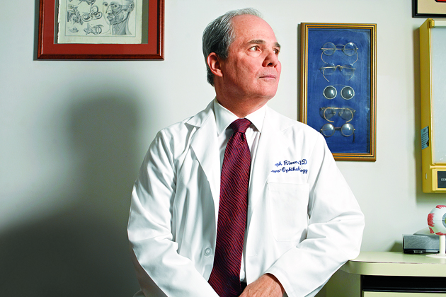
The 50 or so people who have received artificial retinal implants say they can now differentiate walls from windows, detect whether a computer monitor is on, point to the location of a person standing silently in a room and sort dark socks from white ones. Such achievements would represent a major advance in accomplishing what long has seemed beyond hope: restoring sight to the sightless. But some of these cases involve people who had some vision before the implant, making it difficult to know if the devices improved their vision, says neurologist and ophthalmologist Joseph Rizzo, director of neuro-ophthalmology at the Massachusetts Eye and Ear Infirmary and director of the Center for Innovative Visual Rehabilitation at the VA Boston Health Care System.
“It is utterly clear that people who have been blind for decades can see something when you stimulate the retina,” says Rizzo, who in the late 1980s was the first scientist to receive funding for work on an artificial retina. “But there is no evidence that the vision we’re able to create at this point provides enough detail to really make a difference in someone’s life.”
And so, more electrodes. The first generation of artificial retinas, created in the past decade, had as few as 16, while the most advanced current devices have 64. Each electrical contact stimulates cells in the eye—so additional electrodes mean more stimulation and, perhaps, better vision. Typically, the electrodes are embedded in a substrate that is many times thinner than a human hair. This tiny electronic component can be tacked to the front of the retina or slid behind it. Now Rizzo and others are developing prototypes that pack as many as 200 electrodes onto the device. “With 200, people may be able to find the sidewalk and see cars, so they can safely navigate in an unfamiliar environment without a cane or a guide dog,” says Rizzo, who predicts the device will be ready for clinical trials as early as 2011. “Being able to do those things would be a stunningly large accomplishment.”
But if 200 electrodes are good, 1,000 could be much better, suggests Mark S. Humayun, a biomedical engineer and ophthalmologist who serves as associate director of research at the Doheny Retina Institute at the University of Southern California. Humayun expects to produce a 1,000-electrode artificial retina within five years and thinks such a device will enable formerly blind people to read and recognize faces. With each additional electrode, however, comes one more microscopic hole through the chip—a potential source of failure if it provides an opening for the corrosive saltwater of the eye. Electrodes also generate heat, which can burn the eye, and having a denser array increases “cross talk” among the electrodes that may interfere with signals to the optic nerve and brain.
Even surmounting those obstacles will take this technology only so far. Being able to stimulate retinal cells in a way the brain can fully comprehend is the ultimate goal, and although Rizzo thinks scientists will someday find their way across, that could take another two decades. Fortunately, people already losing sight probably won’t have to wait that long for vision repair advances. Gene therapy—once considered a potentially all-powerful therapeutic tool but then widely viewed as generally ineffective and possibly dangerous—has shown great promise in treating one rare eye disease and could be applied more broadly as more genetic targets are identified. New drugs, including one now used effectively against age-related macular degeneration—which affects an estimated 10 million people in the United States—may provide another way to attack vision loss. And work on reprogramming stem cells, though still at an early stage, might eventually provide yet another avenue for re-creating retinal cells and restoring sight. This steady drumbeat of breakthroughs during the past five years has led scientists to hope that treating and even curing devastating eye diseases involving the retina may now be within reach.
THE RETINA IS WHERE SIGHT BEGINS. About the size of a half dollar, this multilayered, light-sensitive tissue lining the back of the eye is crowded with 150 million nerve cells of some 50 different types. The retina forms from brain tissue during embryonic development and is the only conduit of information from the eye to the visual area of the brain.
For someone to see an object, reflected light must pass through the pupil. By growing larger or smaller, the pupil regulates how much light is projected onto the retina. The retina is dense with two types of photoreceptors: cones, which perceive color and fine detail, and rods, which detect fast changes in motion and are responsible for peripheral and night vision. The rods and cones absorb light coming in through the pupil and produce biochemical messages, which are converted to electrical impulses that travel along the optic nerve to the brain, where the signals are converted to images.
Of the many sight-destroying diseases affecting the retina, age-related macular degeneration, or AMD, is the most common, and until four years ago, a diagnosis of AMD meant certain and complete blindness. But the drug Lucentis, approved in 2006 by the Food and Drug Administration, has revolutionized treatment. Adapted from the cancer drug Avastin, Lucentis prevents vision loss in 90% of people with “wet” AMD, in which abnormal blood vessels grow behind the retina and leak blood and fluid into the eye, causing sudden blindness. “We may not be able to cure the disease yet, but at least we can arrest its progression, which is quite remarkable,” says Paul Sieving, director of the National Eye Institute at the National Institutes of Health in Bethesda.
Lucentis blocks the growth factor VEGF, a signaling protein that leads to abnormal blood vessel growth and leakage; one of the most powerful ways to treat disease would be drugs that home in on specific molecular targets. Researchers are now looking for other proteins involved in retinal diseases and for other drugs that could block their effects.
But there’s another, more direct approach to treating gene-related diseases: replacing a malfunctioning gene with a healthy one. While most attempts at gene therapy have turned out to be ineffective, there are several reasons injecting genes into the eye could succeed, says Katherine A. High, director of the Center for Cellular and Molecular Therapeutics at Children’s Hospital of Philadelphia.
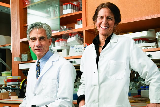
The primary risk in gene therapy is that the immune system will reject either the donated genetic material or the viral vector—a virus that has had its own DNA replaced by the new gene—used to carry the gene into a cell. The body might also mount an autoimmune response that could threaten healthy cells. But since the eye is an “immunoprivileged” site, secluded from the direct blood supply, a harmful immune response is less likely. “Also, several molecules that inhibit the immune response are found in high prevalence in the eye, and it’s geographically segregated from other tissues,” says Jean Bennett, a researcher at the University of Pennsylvania School of Medicinein Philadelphia. “It’s a self-sustaining organ surrounded by pretty tough barriers.” What’s more, the retina, and particularly the tiny macula, are comparatively small, making it easier to deliver a high concentration of viral vectors relative to the number of cells to be treated—a result much more difficult to achieve when replacing genes in large organs.
The eye has yet one more advantage. Because people are born with a full complement of retinal cells, which don’t divide after birth, the healthy gene can be injected directly into the diseased tissue and expected to stay there, continuing to produce the protein that is lacking because of the faulty host gene.
All those factors convinced High, Bennett and others that the eye might indeed be uniquely suitable for gene therapy, and their work on Leber’s congenital amaurosis 2 has produced remarkable breakthroughs. In LCA2, which affects just 80 people in the United States, photoreceptor cells stop functioning and eventually die. The disease is caused by a defect in a single gene, RPE65, that normally produces a protein responsible for processing vitamin A; the retina’s photoreceptors use it to create the visual pigment rhodopsin, which absorbs light entering the eye. Without the RPE65 protein, there’s profound vision loss at birth and total blindness by age 30 or 40.
With just one gene involved and with a window of several decades during which retinal cells are injured but not defunct, LCA2 seemed an ideal testing ground for gene therapy. Bennett and her husband, Albert Maguire, a retina surgeon at Children’s Hospital of Philadelphia, made it work in animals, restoring sight to nearly 60 briards, a sheepdog with a gene that causes severe retinal degeneration similar to that of LCA2. Yet because the human disease is so rare, biotechnology companies had little interest in supporting a trial. So High convinced Children’s Hospital to produce the vectors and test the safety of the gene-transfer therapy.
Twelve people with LCA2, whose ages ranged from 8 to 44, participated in the trial. Before being treated, they all had trouble navigating a dimly lit obstacle course, and several could read nothing on an eye chart and were dependent on others to lead them everywhere. Afterward their vision was partially restored, and within two months a father who hadn’t been able to see his baby’s face was taking the child on neighborhood walks. Road signs and the numbers on cell phones and digital clocks suddenly came into view. The day vision of most subjects improved significantly, and in all subjects, night vision improved as much as 40,000-fold as their retinas became more sensitive to light.
But the most dramatic improvements occurred in four children, ages 8 to 11, who had less damage to their retinas from the progressive disease than did the adults. An eight-year-old who could read only large print on an electronic screen now needs no special visual devices and plays baseball. Another child, who since birth had been able to see only light and shadow, now plays soccer. And all the children navigated the obstacle course more quickly and with fewer errors than before gene therapy. (Not all of the adults improved their performance on the test course.)
Today, more than two years after the first subjects received the gene therapy, their enhanced sight hasn’t waned, and some have even reported ongoing improvements. Although Bennett isn’t sure how long the treatment will be effective, she is already thinking about how gene therapy might be used to treat “a huge list of single-gene defects that destroy the retina’s photoreceptors.” And the breakthrough sets the stage for inserting genes to slow or prevent other eye diseases. “The gene therapy trial for LCA2 has advanced a big area of potential treatment involving 500 identified genes that cause people to lose vision,” says Sieving of the National Eye Institute.
IT’S NO ACCIDENT THAT GENE THERAPY IN THE LCA2 TRIAL WORKED best with the youngest participants. Their retinal cells hadn’t died off altogether and could be revived by the introduction of healthy genes. But that approach won’t help older people, already blind, whose retinal cells will never grow back. For them the best hope may be self-renewing stem cells, which, at least theoretically, can replicate and differentiate themselves into many types of specialized cells—including those in the eye. “By putting a cocktail of stem cells in the eye that will proliferate in the right quantity and have the right characteristics, we can replace missing retinal cells and make the entire retina work again,” says Sieving. “This technology is poised to explode.”
David Gamm, assistant professor of ophthalmology and visual sciences at the University of Wisconsin–Madison, is a leader in that effort. He’s building on a landmark 2007 discovery by other University of Wisconsin scientists, who inserted four genes into an adult skin cell and programmed it back to an embryolike state capable of differentiating into any of the body’s 220 cell types. Gamm has taken those undifferentiated cells—known as induced pluripotent stem cells, or iPS cells—and used a chemical bath of proteins and growth factors to coax them into becoming retinal cells.
This approach has enabled Gamm to observe the entire process of how undifferentiated cells turn into retinal cells, moving from a primitive form to a fully developed retina. Now researchers can study the genes produced by those cells to understand better what goes awry in degenerative retinal diseases.
But perhaps the most exciting possibility is that iPS cells could replace dead or damaged retinal cells. A clinical trial, scheduled to begin soon in the United Kingdom, will test the viability of using embryonic stem cells to replace degenerated retinal pigment epithelium, which nourishes retinal cells, in people with AMD. The stem cells will be placed on an artificial membrane that will be inserted in the back of the eye. If the results are positive, Gamm hopes to test whether iPS cells—which avoid the ethical issues of harvesting stem cells from human embryos—would also work. He has grown retinal cells from both embryonic and induced stem cells and says they appear identical. “Five years from now, there could be clinical trials to replace cells in people with retinal degenerative diseases such as AMD and retinitis pigmentosa,” says Gamm.
RESEARCH ON REPROGRAMMING STEM CELLS, like the work exploring the possibilities of gene therapy and the artificial retina, are all part of a great leap forward in treating eye diseases that not long ago seemed hopeless. When Joan Miller, chief of ophthalmology at the Massachusetts Eye and Ear Infirmary in Boston, finished her fellowship and became a retina specialist in 1991, “no one wanted to treat macular degeneration because it was so depressing—to the patients and physicians,” Miller says. Now, treatment of people with even the most severe form of the disease is actually reversing vision loss, says Miller, who, with renowned vascular biologist Judah Folkman as her mentor, pioneered the use of drug therapy to target the protein VEGF in the eye.
That success has reverberated through the medical community. “It’s exciting that the eye, with all its natural advantages, is leading advances,” says Miller. “Not so long ago, ophthalmology was a surgical subspecialty. Now we use drugs more than surgery, and I have no doubt we’re headed for another revolutionary time in the field, with gene and stem cell therapies.”
Dossier
bostonretinalimplant.org and artificialretina.energy.gov. Ophthalmologists Joseph Rizzo and Mark Humayun have taken different approaches to the development of an artificial retina. Their respective Websites offer videos, interactive models and overviews of their research.
“Age-Dependent Effects of RPE65 Gene Therapy for Leber’s Congenital Amaurosis: A Phase 1 Dose-Escalation Trial,” by Albert M. Maguire et al., The Lancet, Nov. 7, 2009. All 12 trial subjects with an inherited retinal disease had improved sight after therapy to replace a defective gene, paving the way for the therapy to be used on other inherited retinal diseases.
“Gene Therapy for Red-Green Colour Blindness in Adult Primates,” by Katherine Mancuso et al., Nature, Aug. 14, 2009. The authors describe how the gene therapy they gave squirrel monkeys cured them of color blindness—a disorder in which the photoreceptors are not damaged—giving hope that other functions can be added or restored to the eye.