Published On June 20, 2002
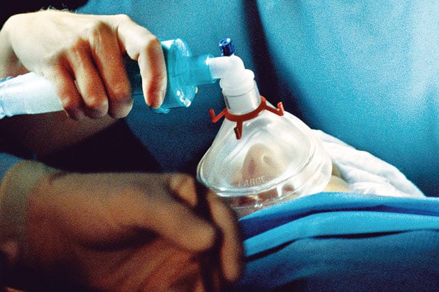
IN A SURGICAL SUITE, A WOMAN IN HER MID-40S LIES MOTIONLESS, her upper and lower body draped with sterile cloth. She has just had her gallbladder removed, and the surgeon is suturing the last of several small abdominal incisions through which he threaded his laparoscopic instruments. Across the table, an anesthesiologist monitors digital readouts showing the patient’s heart rate, blood pressure and other vital signs. When the surgeon finishes suturing, the anesthesiologist cuts the flow of drugs so the patient can find her way back to consciousness.
The case is routine in every way, one of 50,000 every day in the United States in which patients undergo general anesthesia. From long experience, the anesthesiologist expects the woman’s involuntary functions, such as her ability to breathe, to return first; then, she will slowly awaken, first showing small signs of life—a frown, perhaps a tear—and then gradually become aware of her surroundings. Yet now, precisely the opposite is happening. When he speaks, she opens her eyes. He asks her to squeeze his hand, and her firm grasp confirms she is able to recognize commands and carry out high-level cognitive functions. But her brain still is not performing one of its most basic assignments: instructing her lungs to inhale and exhale.
Because the patient’s breathing tube is still in place, the situation is more intriguing than it is alarming. Indeed, data from the pulse oximeter clipped to her middle finger—the device uses light to measure blood oxygen levels—reveals her blood is 100% saturated. Nor is the patient disconcerted by the unusual sequence of events; within a minute, as the anesthesiologist pumps the balloonlike device that forces air into her lungs, her chest begins to rise and fall. But for the anesthesiologist, Emery Brown, a member of the Massachusetts General Hospital’s Department of Anesthesia and Critical Care and an associate professor of anaesthesia at Harvard Medical School, the episode is a stark reminder of how little anesthesiologists really know about the phenomenon to which they have devoted their careers.
During the 160 years since ether was introduced, anesthesiologists have become better and better technicians. General anesthesia is now so routine that people speak of “going under” as casually as they might refer to a stressful but otherwise unexceptional business trip. The risk of a fatal accident during anesthesia has plunged from one in 10,000 cases 36 years ago to just one in 250,000 today.
Still, only now, after long and steady improvements in the practice of anesthesia, are researchers beginning to piece together what happens in the brain of an anesthetized patient. Advances in imaging technology, information processing, molecular biology and other specialties are enabling scientists to map the functions of various brain regions, study the brain’s electrical waves and analyze communications between individual neurons. In the process, a specialty sometimes regarded as almost a trade in the medical hierarchy—an essential service but hardly a hotbed of research—is becoming an arena of cutting-edge inquiry.
At stake is much more than satisfying intellectual curiosity. Grasping how anesthetics affect specific parts of the brain could help researchers develop better-targeted drugs that shut down only certain functions—those involved with feeling or remembering pain, for example—while leaving others unimpaired. According to Ira Rampil, professor and director of clinical research in anesthesiology at Stony Brook University in New York, the current array of anesthetics are reliable tools for putting patients under and bringing them back, and anesthesiologists tend to use much the same procedure for every patient. But with more specialized choices, physicians could tailor drug and dosage to each case. “We could become fine chefs rather than short-order cooks,” Rampil says.
AS EARLY AS 1800, PHYSICIANS IN EUROPE and the United States knew that compounds such as nitrous oxide, or laughing gas, had pain-deadening properties. But it wasn’t until 1842 that a physician from Georgia, Crawford W. Long, used ether to anesthetize a patient who was having a tumor removed from his neck. Four years later, in the first clinical demonstration of anesthesia, a packed gallery at the Massachusetts General Hospital watched William T. G. Morton, a Boston dentist who had used ether to make tooth extractions less painful, administer ether vapors to a 20-year-old printer named Edward Gilbert Abbott. John Collins Warren, the hospital’s chief surgeon, then removed a vascular malformation from Abbott’s neck. When he finished, Warren proclaimed, “Gentlemen, this is no humbug.” Word of the procedure, which the physician and poet Oliver Wendell Holmes dubbed “anesthesia,” quickly spread across America and Europe.
The advance was sorely needed. Before anesthesia, even minor surgery was nightmarish, and patients who survived often carried psychological scars for the rest of their days. Moreover, many procedures that are now routine, such as appendectomies, were all but impossible to perform, because there was no way to keep a patient from writhing in agony when the knife cut into the body, and chances were high that he would die from shock.
As anesthesia became widely used, a few facts emerged. It turned out that, appearances to the contrary, going under has little to do with sleep. In even the deepest sleep, we toss and turn, feel pain and can be roused by a noise or a shake. An anesthetized patient is in a sort of suspended animation, insensible to noise or any other disturbance. Though the heart continues to beat, the patient has no awareness of a scalpel slicing into flesh, an organ being pushed aside, a tumor being excised. Somehow, propofol and other widely used anesthetic drugs interfere with the brain’s ability to send and receive messages.
“You can’t be aroused from anesthesia under even the most noxious stimulations, because the cognitive ability of the brain has been turned off,” says Steven Shafer, professor of anesthesiology at Stanford University. “There’s nothing else in pharmacology that’s quite like this. We don’t have drugs that turn off the liver or other organs, and yet we are able to turn off the cognitive ability of the brain—and then turn it on again.”
During much of the twentieth century, it was thought that anesthetics shut down the entire brain, akin to turning out a light and leaving a room in total darkness. But recently that theory has given way to a more complex picture of a room divided into cubicles, some of which remain lit while others go dark. Even now, though, nothing is known for certain.
HOWEVER ANESTHESIA WORKS, A MAJOR BENEFIT FOR PATIENTS—in addition, of course, to avoiding pain—is remembering nothing afterward. “What patients want is oblivion,” says Rampil. Sometimes, though, despite anesthesia, patients are aware of what they’re experiencing and may be left with traumatizing memories. And though such cases are rare, the frequent use of general anesthesia means that even a rate of one or two per 1,000 cases results in as many as 40,000 U.S. patients each year who suffer from recall.
Preventing recall is difficult because each patient has a different tolerance for anesthesia. A level of drug that renders one patient unaware (and thus safeguarded against recall) might leave another insufficiently sedated. Most anesthesiologists have tended to err on the side of caution, administering ample doses to ensure forgetfulness. But recently, they’ve been able to employ a more precise approach, using electroencephalograms (EEGs) to monitor the brain’s electrical activity under anesthesia.
The EEG is, medically speaking, an ancient technology, introduced more than 125 years ago by a British physician named Richard Caton. But a device developed in the 1990s by Aspect Medical Systems, a Massachusetts company, translates EEG readings into a single number called a BIS Value. On the bispectral index’s scale of zero to 100, the highest BIS numbers represent degrees of full consciousness. A fully alert patient might have a BIS reading of 94; as the level of anesthetic increases, the number steadily falls. At a BIS of 60 or lower, the probability of a patient being subject to recall is reduced by as much as 80%.
An EEG can also produce graphs showing the amplitude and frequency of electrical activity in the brain. With patients who are alert or even in conventional sleep, the tracings on the graphs (the brain waves) seem to make little sense. The points charted are packed together and dart up and down in no apparent pattern. That’s because a brain’s normal electrical activity is ever changing, rising and falling as the barrage of sensory impulses is processed into thought and action.
Under anesthesia, though, a striking pattern emerges. As more drugs are administered, the frequency of activity drops, and the spikes of the brain waves are higher and spread farther apart. Eventually, as the subject enters a state of deep anesthesia, the scattered waves elongate and reveal a series of long, slow ocean swells. Yet as with so much else associated with anesthesia, it isn’t certain just why the swells occur.
To begin to unlock that secret, Brown and his research team have used both EEGs and functional magnetic resonance imaging (fMRI). Unlike conventional MRIs, which yield detailed pictures of body parts, fMRIs map areas of brain activity by tracking blood flow through thousands of tiny veins and arteries. Oxygenated blood (carrying oxygen to the cells) and deoxygenated blood (returning to the heart and lungs) have different magnetic properties. When captured by fMRI, those characteristics translate into intricate pictures of brain activity.
In Brown’s experiments, subjects are slipped into the tunnel of a machine, wearing nonmagnetic helmets with electrical receptors that feed signals from the brain to an EEG monitor. They are put under anesthesia in stages, with detailed fMRIs taken during each stage. Blood increases and decreases relative to the amount of electrical activity going on in a particular area, showing which parts of the brain are active under anesthesia.
Because fMRI images take several minutes to produce, volunteers must be anesthetized very slowly. That means a breathing tube must be in place while a subject is still fully awake. Because most people would gag on the tube, Brown’s team has sought out volunteers with tracheostomies, which provide a built-in external connection for the tube. In more than a year, studies have been conducted on only four volunteers. Even so, initial results are promising.
EVERY THOUGHT TRAVELS AN ELABORATE PATHWAY through the brain. When the brain responds to a sight or sound, sensory impressions move directly from the optic or auditory nerve to the thalamus, the clearinghouse for sensory information. They then go to the cortex, where the electrical impulse carried by the brain’s circuitry is processed into a conscious thought—say, associating a certain sound with a particular musical instrument.
Subjects in Brown’s experiments are asked to respond to auditory stimuli. The volunteer presses a button to indicate whether a tone is high or low pitched. With the subject under a light dose of propofol, the EEG graph still shows the choppy up and down of active thought, and the subject is sufficiently alert to respond to 90% of the tones. With a moderate dose, brain waves begin to elongate, and the subject can respond to only two out of three tones. Under a heavy dose, the brain waves form long swells, and there’s no response to the tones.
Yet even then, the brain continues to process sound. The fMRI images show auditory information making its way through the thalamus to the auditory cortex. In other words, the brain still appears to recognize sound as sound, even though the subject is unconscious and will have no memory of hearing it.
More tests will be needed to establish a method to gauge how far into the cortex auditory signals advance before being snuffed out. Then, Brown and his team hope to conduct tests that substitute pain for sound. If they ultimately locate the specific regions at which anesthesia deadens pain, the next step could be to develop a drug that affects just those parts of the brain.
Brain imaging with fMRIs goes a step beyond EEGs in determining what happens when a patient is anesthetized. Yet even fMRIs can’t measure activity at its most basic level—individual neurons transmitting information.
IN ORDER TO COMMUNICATE WITH ONE ANOTHER, brain cells release chemicals that bridge the gap between cells. Some of these agents, called neurotransmitters, are “excitatory”—that is, they pass along a call to action, while others inhibit action. “Anesthetics seem to decrease the activity of excitatory synapses, and at the same time magnify the effects of the inhibitory synapses,” says Neil Harrison, professor of pharmacology in anesthesiology at Cornell University’s Weill Medical College in New York City. The brain’s primary inhibitory transmitter is known as gamma-aminobutyric acid (GABA), and Harrison thinks that anesthetics somehow enhance the ability of GABA receptors to keep messages from making their way through the brain.
During the 1990s, to test this theory, Harrison and others added brainlike GABA receptors to human kidney cells (which have no such receptors). Then they applied GABA to the cells to imitate cell-to-cell communication and recorded the minute electrical charges that resulted. When anesthetic drugs were introduced to the cells, the electrical signals caused by GABA became stronger. Next, the researchers genetically modified the receptors so that they continued to respond to GABA but now no longer responded to anesthetics. It was a landmark finding because it supported the idea that anesthetic drugs indeed target specific receptors.
Several years later, competing researchers in Zurich and Pittsburgh bred mice with altered GABA receptors, and some of the animals proved to be up to 30 times more resistant to anesthesia than normal mice would be. Now scientists are trying to determine exactly how anesthetic drugs bind to the receptors. Unlocking that puzzle might lead to more targeted anesthetics as well as to drugs that could quickly reverse the effects of anesthesia.
As an undergraduate at Cambridge University during the early 1980s, Harrison was lured to the study of anesthesiology by how little was known. In a pharmacology course, professors provided detailed descriptions of how antibiotics and other drugs did their work. “But in the lecture on anesthetics, the whole story was so vague it didn’t make sense,” says Harrison. “The whole thing struck me as unsatisfying.”
For Harrison and others, that dissatisfaction is giving way to a sense of hope. “We have been able to figure out with great precision how the vast majority of drugs work,” says Stanford’s Steven Shafer. “I can’t think of any other conventional pharmaceutical that’s more than 10 years old for which we don’t know the mechanism of action.” Now, after 160 years, researchers may finally be on the road to solving what Shafer calls “the oldest unsolved mystery in pharmacology.
Dossier
“A Primer for EEG Signal Processing in Anesthesia,” by Ira J. Rampil,Anesthesia, October 1998. A definitive if highly technical account of BIS readings and other uses of EEG in anesthesiology.
“The Effects of Anesthetics on Brain Activity and Cognitive Function,” by Wolfgang Heinke and Stefan Koelsch, Current Opinion in Anaesthesiology, December 2005. An excellent overview of recent work, from EEGs to neuroimaging.
“Consciousness Unbound,” by George A. Mashour, Anesthesiology, February 2004. A reflection on the various “cognitive binding” theories of consciousness, and their implications for anesthesia.