Published On January 24, 2018
IN 2016 SEVERAL TOP cancer experts convened at a forum sponsored by Partners HealthCare to produce a report on the technologies that were most likely to transform cancer care during the next decade. Daniel Haber, director of the Massachusetts General Cancer Center, remembers the group talking about CRISPR gene editing, artificial intelligence to improve diagnosis and treatment plans, and immunotherapy techniques that incite the immune system to fight tumors. “At the end of the conversation someone said, ‘What about nanotechnology?’” Haber says.
It’s an open question. Among those experts, the consensus was that while nanotechnology, which in cancer treatment uses vanishingly minuscule particles to launch sneak attacks on tumors, may have promise for the future, that future has always felt just out of reach. Since the mid-1990s, when the first of a handful of nanotech cancer drugs hit the market, several have become standard parts of chemotherapy. Yet this approach to battling cancer has never proved quite as revolutionary as it was supposed to be. “For decades, everyone’s been talking about this exciting research, and there’s no doubt they’re making some really cool particles,” Haber says. “But we’re still waiting for the breakthrough moment when we can say, ‘This nanotech has a real impact in the clinic.’”
Now that moment may finally be coming into view. At a time when gene therapy has been revived as a potent form of cancer treatment, a new approach would use nanoparticles, rather than viruses, to deliver strands of DNA or RNA to tumors. Other nanoparticles in development could be injected to rev up the immune system’s ability to attack malignant cells. Researchers are experimenting with two ways to use gold nanoparticles—in one case, to reduce the collateral damage caused during radiation therapy while increasing effectiveness, and in another, as the targets of laser beams to help kill tumor cells. Iron-based nanoparticles, meanwhile, can be activated by magnetic fields to generate tumor-scorching heat.
At one hub of innovation, MIT’s new Marble Center for Cancer Nanomedicine, researchers have taken aim at ovarian cancer with wide-ranging approaches, crafting nanoparticles designed to diagnose or treat the deadly disease. In their most intriguing work, they’re collaborating on something new: a “theranostic” nanoparticle that can both diagnose and attack ovarian tumors. Such a potent particle would go well beyond the limits of today’s cancer nanomedicines—and could finally demonstrate what can happen when cancer researchers think small.
THE FIRST NANO CANCER drug approved by the Food and Drug Administration in 1995 was designed to make an existing treatment better, and the drug it would work with was a commonly prescribed chemotherapy, doxorubicin. Although doxorubicin can be effective at killing tumor cells, its noxious impact can hurt healthy tissues as well. Too large a dose can cause congestive heart failure, among other side effects, while a small dose may be overwhelmed by immune cells, which consider doxorubicin a foreign invader.
Yechezkel Barenholz, who was working at the Hebrew University Hadassah Medical School in Israel, and his colleagues came up with a nano packaging trick that addressed those problems. To get an idea of the scale at which they were working, consider that a human hair is about 100,000 nanometers wide. Barenholz’s team put doxorubicin inside 100-nanometer particles made of lipid (fat) molecules, decorated on the surface with a polymer that attracted water molecules. That allowed each nanoparticle, packed with medication, to circulate in the blood in a surrounding cloud of water that shielded it from immune cells.
The clever packaging also reduced toxic side effects by using a quirk of tumor anatomy. In its rush to grow, cancer forms tangles of blood vessels, but those slapdash vessels are leaky. Barenholz’s nanoparticles generally stayed in the bloodstream and avoided the heart and other organs, but when they reached the leaky blood vessels in the tumor, they slipped through those holes. When the particles reached the tumor itself, they released their chemo payload in a process related to that particular tumor cell’s metabolism. This passive targeting system meant that a low dose of the drug could make a strong impact on tumors, yet with few side effects.
“When we started, no one believed it would work,” says Barenholz, now a professor emeritus of biochemistry and molecular biology at the Hebrew University of Jerusalem. But in 1995, after successful clinical trials, the FDA approved the nanodrug Doxil for treating AIDS-related Kaposi sarcoma; later, the agency extended its approved use to ovarian cancer and multiple myeloma, and in Europe doctors use it to fight breast cancer.
Like Doxil, all other existing cancer nanomedicines use specially engineered nanoparticles to transport an existing chemotherapy agent. And by some metrics, these first-generation nanodrugs have done well. They are widely prescribed and have been shown to limit toxic side effects.
Studies have found, however, that Doxil doesn’t perform significantly better than the original doxorubicin drug at slowing cancer’s advance or in prolonging patients’ lives. Some analyses have questioned whether the passive targeting strategy, which depends on the circulating medication slipping through the unique apertures of a tumor, is effective, noting that the blood vessels that spring up around tumors may not be as porous as originally thought, and therefore may prevent large quantities of the nanodrug from reaching its target. While some researchers have tried to boost efficacy through new ways to administer the drug, such as in conjunction with focused ultrasound, others have looked to completely different approaches.
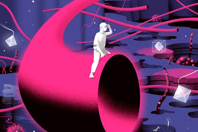
Illustration by Señor Salme.
TO MAKE DEEPER INROADS against cancer, nanotechnology may need to do more than simply miniaturize standard treatments. At the Marble Center for Cancer Nanomedicine, three research groups are working on particles that take more complex approaches to targeting tumors and delivering a wide variety of substances, including new kinds of diagnostic agents as well as cancer-killing therapies. Many of these efforts are focused on ovarian cancer, which is particularly deadly, with only 47% of those with the disease alive five years after diagnosis.
One group is led by Angela Belcher, a relative newcomer to medical research. After years of breaking ground on nanomaterials for batteries and solar panels, in 2010 she joined MIT’s Koch Institute for Integrative Cancer Research and threw herself into solving the riddles of ovarian cancer. Belcher’s background, she feels, gives her a clear perspective on human anatomy and its quirks. “I’m a materials scientist, so I look at everything as a material,” she says. “To me, all problems have material solutions.”
Belcher, a professor in two MIT departments—materials science and biological engineering—believes there’s a misperception regarding nanotech. “A lot of people think the great thing is that it’s small,” she says. “But what’s really great is that when a material gets that small, its properties change.” In that tiny world, objects have different optical, magnetic, electrical and mechanical attributes. Belcher explains that nanotech researchers can “tune” a material to get the properties and the outcomes they want.
Belcher’s system uses nanoparticles as an imaging system that can help surgeons find tiny bits of residual tumor. “We’ve known for years that how well a patient does is directly related to the amount of tumor that the surgeon removes and the amount that’s left behind,” says Michael Birrer, who began working with Belcher when he was head of medical gynecologic oncology at Massachusetts General Hospital. (He’s now director of the Comprehensive Cancer Center at the University of Alabama at Birmingham.) Tumor cells that aren’t taken out can grow and spread, often with deadly consequences. With Belcher’s nanoparticles, surgeons should be able to spot and remove tiny clusters of just a few cells, even when they’re hidden behind other organs, thus preventing those seeds from growing into major malignancies. “Theoretically, we may be able to cure some patients,” Birrer says. “This is some of the most exciting work I’ve ever done.”
The crucial element is a carbon nanotube, a hollow structure made of sheets of carbon only one atom thick. At that scale, when the nanotubes are hit by near-infrared light with wavelengths of about 800 to 1,400 nanometers, they naturally fluoresce. That fluorescence isn’t visible to the naked eye, but it can be easily recorded with optical equipment.
Introduced during tumor removal surgery, the nanotubes could light the way for surgeons. In a recent study using mice, a virus that binds to the outside of ovarian cancer cells was used to deliver nanotubes to the tumor sites. After the bulk of the ovarian tumors had been removed, the team projected near-infrared light onto the surgical site. The nanotubes illuminated minuscule tumor fragments—some as small as half a millimeter in diameter—that the surgeons then removed. “We increased median survival time in animals by 40%, which gives us enough evidence to go on to human clinical trials,” Belcher says. The team has already submitted a request to the FDA to conduct a small initial study in people.
Belcher hopes that the nanotubes might ultimately serve an additional function, as part of a noninvasive imaging system to screen women for ovarian cancer. The near-infrared light that causes the nanotubes to fluoresce can penetrate about eight centimeters into human tissue, so physicians could potentially shine the light through skin and flesh to look for fluorescence from nanotubes signaling the presence of cancer cells. Women whose ovarian cancer is detected before it has spread have a much better five-year survival rate of 93%. “That’s the work I’m most excited about,” Belcher says. “I want to find those early-stage tumors, and I want to increase the survival of patients with ovarian cancer.”
THE LAB OF SANGEETA BHATIA, director of the Marble Center, is also working on a diagnostic technique for ovarian cancer, but this one relies on chemistry. The researchers have devised a nanosensor that breaks apart in the presence of ovarian tumor cells and sheds fragments of itself. Those fragments are then filtered out through the kidneys and can be detected in a urine test.
The nanosensors accumulate in the tumor through a turbocharged variation of the targeting mechanism that first-generation cancer nanodrugs employ. Once the sensors have traveled into a tumor’s leaky blood vessels, they use special targeting molecules to bind to receptors on the surface of blood vessel cells. That activates a cellular process that whisks the nanosensors inside the tumor’s outer shell, where they can do their work. The sensors are particularly helpful because they respond to a specific enzyme that a tumor needs to grow blood vessels and to remodel neighboring tissue, steps that enable the tumor to spread. The enzyme “isn’t just a random by-product of a tumor—it’s something that can serve as a marker for growth and malignancy,” says Ester Kwon, a former postdoctoral researcher in Bhatia’s lab.
Small molecules called peptides on the nanosensor’s surface react chemically with the enzyme, causing fragments of the peptides to break off. Those fragments float away, are captured by the body’s filtration systems, and wind up in urine—in which they can be detected even at minute levels.
The nanosensors aren’t yet ready to be tried in people. But in mouse studies, the sensors detected tumors smaller than five millimeters.
Like Belcher’s nanotubes, this diagnostic tool could also be used as a screening method for ovarian cancer. Today, women typically don’t get checked until they’re exhibiting symptoms, which can range from abdominal pain and indigestion to constipation, diarrhea and vaginal bleeding. Moreover, current diagnostic tests aren’t very sensitive. The average tumor detected by ultrasound measures about five centimeters in diameter, and the biomarkers used in blood tests enable oncologists to detect tumors as small as one centimeter. Researchers estimate that it can take as long as 10 years for a tumor to grow that large. Bhatia’s technology could allow doctors to detect the cancer and provide treatment months or even years earlier than they do today. That difference could be lifesaving.

Illustration by Paulo Campos.
A THIRD INNOVATION FROM the Marble Center comes with an arresting moniker: the gobstopper, named after the multilayered hard candy. It’s the result of an effort by Paula Hammond, who heads MIT’s chemical engineering department, to perfect a process of layer-by-layer assembly that enables nanoparticles to carry several drugs between their strata. The gobstopper can then sneak those therapeutic agents past the body’s defense systems and deliver them to an ovarian cancer tumor.
The outer layer of the gobstopper is a stealth layer; much as in the technique used by Doxil and other first-generation nanodrugs, its surface attracts water molecules, so the particle can avoid notice by the body’s patrolling immune cells. Its outer coating also has a negative electrical charge that repels the negatively charged immune cells.
When the nanoparticle reaches a tumor, its middle layer releases an RNA molecule that weakens the tumor’s defenses by turning off certain cancer-promoting genes. That assault sets up the tumor for a final blow when the nanoparticle’s core releases a chemotherapy agent. “By building particles layer by layer, we’ve already made two compartments for drugs,” Hammond says. “And we can get fancier.”
The modular nature of Hammond’s nanoparticles might also benefit either Belcher’s light-responsive nanotubes or Bhatia’s fragment-shedding nanosensors, by packing them into a layered particle that contains a chemo drug at its core. Hammond has ongoing investigations with both of those labs, work that could lead to a theranostic nanoparticle that simultaneously identifies ovarian tumors and hits them hard.
Such a combination, Hammond says, could yield rich information for oncologists. “With the nanotubes, we’d have a way of optically imaging the location of tiny tumors and treating them on the spot,” she says. “Then with the second method, we can detect tumor activity with enzymatic activity, and can monitor the effects of treatment over time.”
With the enzymatic detection method, says Bhatia, urine tests could also be used to track a patient’s progress. If the signal decreased, the oncologist would know the tumor was shrinking. “You could assess the patient’s response to treatment, alter the regimen if needed, and monitor for any chance of early recurrence,” she says. That adaptive approach to treatment might offer patients relief from chemotherapy’s harsh side effects. “We’re trying to increase both survival and quality of life,” Bhatia adds.
The Marble Center researchers, as well as others in this field of cancer treatment, are well aware that these medicines are sometimes characterized as futuristic solutions that are always about 10 years away. It’s true, moreover, that these potential advances aren’t yet close to routine clinical use—and some might not get there at all. But with truly innovative ideas now emerging from the nanoworld—ideas that don’t just scale down existing treatments but take entirely new approaches—they see potential for real breakthroughs that will save patients’ lives. The revolution in cancer care just might be nano-sized.
Dossier
“Mechanisms and Barriers in Cancer Nanomedicine: Addressing Challenges, Looking for Solutions,” by Thomas J. Anchordoquy et al., ACS Nano, January 2017. The result of a workshop involving several dozen leading experts on cancer nanomedicine, this paper discusses the limitations of current cancer nanodrugs and the research necessary to advance the field.
“The Evolving Landscape of Drug Products Containing Nanomaterials in the United States,” by Sheetal R. D’Mello et al., Nature Nanotechnology, June 2017. Researchers at the Food and Drug Administration examine data from more than 350 nanomedicines submitted for approval since the 1970s and identify trends.
“Plenty of Room at the Bottom,” by Richard P. Feynman, lecture delivered at a meeting of the American Physical Society at the California Institute of Technology, December 1959. This lecture by the theoretical physicist Richard Feynman is often cited as the origin of nanotechnology. He challenged scientists to make machines and products that could interact with the world on the atomic scale.