Published On May 3, 2011
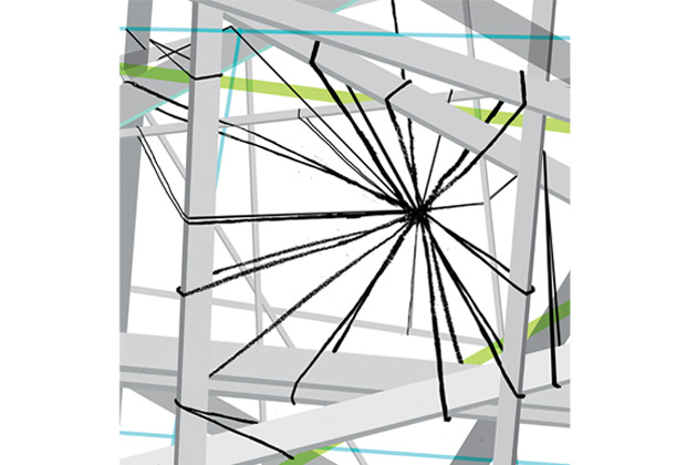
IN 1888, WHEN THE SPANISH ANATOMIST SANTIAGO RAMÓN Y CAJAL began almost obsessively drawing every kind of brain tissue he could obtain—from humans, rabbits, pigs, dogs, fish, frogs, mice and other creatures—he depicted many hundreds of glia, a newly discovered cell type whose fine tendrils radiating in all directions from a small cell body fascinated (and, as an artist, delighted) him. Glia were thought to serve chiefly as “glue” (glia is Greek for “glue”), cementing the brain’s other cells, but to Ramón y Cajal they looked like spiders, and he dubbed them células aracneiformes—spider cells.
Yet it was a second type of brain cell, the neuron, that ultimately captivated him. Neurons had been discovered only a few decades earlier, and anatomists were still trying to puzzle out how they functioned. Studying their form, Ramón y Cajal began to decipher how they communicated, and he developed a theory that would become known as the neuron doctrine, whose basic tenets continue to inform neuroscience. Ramón y Cajal surmised, correctly, that neurons relay electrical signals from one cell to the next throughout the nervous system. The signals are called action potentials, and they travel in one direction only, from the cell’s rootlike dendrites to its axon, a long, slender central section that ends at a tiny gap, the synapse, across which lies the dendrite of the next neuron.
The electrical activity spurs the release of chemicals called neurotransmitters. This happens when gated calcium ion channels open and calcium flows into the presynaptic neuron, triggering the release of neurotransmitters. The receiving, or postsynaptic, neuron has specialized protein receptors in its dendrites that accept the neurotransmitters, causing the neuron’s voltage to drop, firing an impulse down its axon. This in turn releases neurotransmitters from the second neuron and transmits them across the synapse to the next neuron’s dendrites, all at speeds as fast as 200 miles per hour.
Glia, in contrast, were considered incapable of communicating with one another, let alone with neurons. Besides holding neurons in place, glia seemed to fulfill just a few mundane housekeeping tasks, all in the service of the more exalted neurons. This apparently subservient role meant that, for more than a century, glia have gotten little respect and drawn scant attention from scientists, who, in any event, lacked the tools to decipher what these brain cells are doing.
Researchers have long been able to measure the electrical potential of neurons. But glia communicate more subtly, so even though they make up approximately half the volume of the human brain, researchers couldn’t gauge the impact of their role. Only gradually, with the development of sophisticated imaging techniques for calcium, which glia use to communicate, has it become clear that glia handle far more complex jobs than previously thought. Even their housekeeping functions, it turns out, are extremely important to the nervous system’s well-being.
Now, in fact, a growing cadre of neuroscientists is beginning to realize that glia are as fundamental a part of neural circuitry as neurons, probably involved in the development of the brain’s wiring and in learning and memory, blood and energy supply, neurotransmitter balance and more. And as these scientists start to more fully understand the connections between glia and neurons, they anticipate that glia could become important targets of therapy for many disorders of the nervous system, from brain cancers and spinal cord injuries to such neurological diseases as multiple sclerosis and Parkinson’s.
GLIA COME IN FOUR FLAVORS. Astrocytes, the spiderlike cells that intrigued Ramón y Cajal, are found in the brain, their fine branching tendrils wrapped delicately around neurons. Each astrocyte envelops multiple neurons, forming a “domain” distinct from the domains of other astrocytes—in the human brain, one astrocyte’s domain is estimated to connect to tens of thousands of synapses. These “spider cells” also communicate closely with blood vessels in the brain, signaling them to increase blood flow when neurons need additional oxygen or glucose. It’s thought that astrocytes convert glucose into lactate, which is then sent to neurons and converted to pyruvate, a necessary component of the cell’s energy molecule ATP. Two other glia types, oligodendrocytes and Schwann cells, enclose the axons of neurons in myelin, a protective sheath much like the insulation around electrical wires. Microglia, the fourth type, are the nervous system’s immune cells, responding to injury, cleaning up dead cells and debris, and playing a role—whether helpful or harmful is not yet clear—in such neurodegenerative disorders as Alzheimer’s and Parkinson’s. (Microglia are distinct from the other types of glia and generally are not involved in the functions of those other cells. Their numbers also fluctuate widely.)
In 1991, Richard Robitaille, a professor of physiology at the University of Montreal, Quebec, working with his mentor Milton Charlton, used a fluorescent calcium indicator and a fluorescent microscope to study the action potential in a piece of frog muscle and in the motor nerve that told the muscle when to contract. Robitaille fired an electrical stimulator to induce an action potential in the motor nerve, expecting to see calcium flow into the presynaptic neuron, which would indicate that the neurotransmitters were about to release. Instead, through his microscope, he saw cells on top of the neuromuscular junction—where the neuron contacts the muscle—begin to fluoresce, indicating an increase in calcium ions in those cells, which he recognized as glia. At the time, Robitaille and his colleagues thought this was simply a nuisance because the glia were getting in the way of their seeing the function of the synapse. So the scientists shut down the experiment and went home, feeling defeated.
But the more they thought about what they had seen, the more surprising it seemed. “The next day,” Robitaille says, “we asked each other, ‘Did you know glial cells were sensitive to synaptic activity?’ None of us did. We all thought glia were just useless, stupid cells.” Yet this experiment suggested the opposite—that the movement of calcium into glia might mean they played some kind of significant role. Robitaille had been interested in studying neurons, but this accident shifted the focus of his career, and he now studies glia.
Other scientists, in fact, had already begun to decipher what was causing the action that Robitaille observed. In 1990, Ann Cornell-Bell and her colleagues, then at Yale University, had published a paper showing that glia receive signals from neurons, using special channels in the glial cell membrane that are opened by a neurotransmitter called glutamate, causing calcium ions to flow into the cell. The study also demonstrated that the calcium rise in one astrocyte causes a similar surge in nearby astrocytes, creating a wave of increased calcium levels, which suggests that glia not only receive signals from neurons but also pass information among themselves.
Then, in 1994, Maiken Nedergaard, a neurophysiologist at New York Medical College in Valhalla, N.Y., showed that the increase in astrocytes’ calcium levels was followed by a rise in calcium in nearby neurons as well. So it turned out that glia could also talk to neurons, and Nedergaard concluded that this communication happened across gap junctions, the small spaces that connect astrocytes and neurons. That same year, Philip Haydon, then at Iowa State University in Ames, revealed more of this emerging picture, showing that glia release glutamate, which then docks at a specialized receptor on a nearby neuron and raises calcium levels in the neuron.
YET EVEN AS THEY LEARNED MORE ABOUT GLIA, until about 15 years ago most neuroscientists thought they played only a supportive role. Now, with advances in calcium imaging technology and deeper research, that view has been slowly changing. “We’re still just beginning to learn how astrocytic calcium signaling actually works,” says Michael Halassa, a psychiatric resident at Massachusetts General Hospital and a glia researcher who has worked in Haydon’s lab. “But it seems to be a very complex language.”
Astrocytic calcium signaling seems to differ from the action-potential signaling of neurons. Glial signaling occurs in degrees, instead of being all or none, and occurs slowly—with the signal moving within a cell and from one cell to another in seconds or minutes, compared with the milliseconds of an action potential.
Robitaille says recent evidence proves that there is a subtle interplay between glia and neurons and that glia, like neurons, can parse patterns of information at the synapse (by taking up some of the neuron’s neurotransmitters), process them (by using increasing levels of intracellular calcium for signaling) and influence the outcome of synaptic communication based on what they’ve learned (by releasing transmitters of their own into the synapse to affect synaptic activity). If the synapse acts as a dimmer switch that tunes the nervous system, “it seems that glia have their hands on the switch and can intelligently increase or decrease the light,” says Robitaille. He and his colleagues speculate that glia are constantly detecting, decoding and analyzing what is happening at the synapse, and using that information to control the mechanisms that affect how the synapse changes.
If this is correct, it could have profound consequences for the way neuroscientists look at the brain. Given that glia make up about half the volume of the brain and that each astrocyte can make tens of thousands of connections to neurons and to other astrocytes, some researchers estimate that the information processing going on in the brain may be of an order of magnitude more complex than most people have believed.
Glia, it seems, are also involved in synaptic plasticity, a process that determines when and where new synapses form. Typically synapses are created when we learn or form memories, as well as when the brain develops in utero and throughout childhood. In 2005, Ben Barres, a professor of neurobiology at Stanford University, and his colleagues found that thrombospondin, a protein active during brain development and after injury, is also released by astrocytes to activate synapse formation. More recent research suggests that astrocytes secrete other signals that promote synapse formation and function, although the precise nature of glial cells’ role in synaptic plasticity is still unclear.
AN IMPORTANT TECHNOLOGICAL KEY TO UNDERSTANDING GLIAL CELLS is the recently developed ability to manipulate the genetics of individual cells. This allows researchers to change a single gene and then observe how the change affects the glial cell’s behavior. Haydon and his colleagues have used this technique to elucidate the role of astrocytes in regulating sleep. They altered a gene in mice that controls astrocytes’ production of a chemical called adenosine, whose levels normally rise during wakefulness and decline during sleep. The gene alteration caused adenosine levels in the brain to drop, preventing the sleep homeostat from functioning. (The sleep homeostat normally encourages sleep based on the number of hours a human or an animal has been awake.) They concluded that if reducing the astrocytes’ production of adenosine was significant enough to wreck the homeostat, then astrocytes must be significantly involved in regulating the sleep cycle—a discovery that suggests astrocytes can regulate behavior. Indeed, glial involvement in sleep regulation could imply that the cells play a role in psychiatric disorders, many of which involve sleep problems.
Other brain diseases, too, seem likely to involve glia. Haydon and his colleagues are researching the role of glia in epilepsy. “For a long time, we’ve known that in the epileptic brain, astrocytes enter a state called reactive astrocytosis,” he says. “We don’t know what that really means, except that some of the proteins normally associated with glia change, in what we think is a reaction of the glial cell to some kind of neuronal dysfunction.”
Studies also suggest roles for glia in several other neurodegenerative disorders, including Parkinson’s, which is characterized by the death of dopamine-producing neurons in a part of the brain called the substantia nigra, as well as by the formation of aggregates of protein within brain cells. In this case, astrocytes seem to play both helpful and harmful roles. They help preserve neurons by releasing antioxidants and other protective factors and removing toxic molecules from the extracellular space. Yet astrocytes also release cytokines, which promote inflammation that can damage cells, and other molecules that hasten the death of neurons. If researchers can find a way to foster the helpful role of glia in Parkinson’s disease while minimizing the harm they do, that might help them develop a treatment for the disorder.
Finally, glia may play a key role in amyotrophic lateral sclerosis, a fatal disorder in which motor neurons progressively deteriorate, eventually resulting in paralysis. About 1 in 10 cases is hereditary, and in 1993 geneticist Daniel Rosen, then at MGH, and his colleagues, Robert H. Brown of MGH and H. Robert Horvitz of MIT, discovered mutations in a gene that produces an antioxidant enzyme called superoxide dismutase 1, or SOD1, which is one cause of familial ALS. When formed normally, SOD1 scavenges and neutralizes free radicals, oxygen atoms that are corrosive to tissue. Researchers theorized that the presence of mutant SOD1 proteins in motor neurons might become toxic, eventually leading to cell death. And studies have proved that in mice, the SOD1 gene mutation does cause ALS. Further research traced the problem to astrocytes: If the glial cells produce mutant SOD1, astrocytosis results. Although it is not yet clear how this occurs, recent evidence suggests that glutamate may rise to toxic levels in ALS.
Jeffrey Rothstein, a professor of neuroscience at Johns Hopkins University in Baltimore, is studying glutamate transporters with the goal of elucidating the role of astrocytes in ALS and developing treatments.
NOW THAT SCIENTISTS ARE BEGINNING TO REALIZE THAT GLIAL CELLS ARE AN INTEGRAL PART of neural circuitry, a novel and exciting picture of the brain has started to emerge that shows glia in essential partnerships with neurons. Indeed, because of the slower time scale of calcium waves compared with neurons’ instant messages, glia may show their true selves in higher-level cognitive processing. Research is suggesting roles for glia not only in neurotransmitter balance, synapse development and neurological disorders but also in chronic pain, spinal cord injury, depression and more.
“I think what’s coming next,” says Robitaille, “is that we’ll be able to take all the things we know about glia and start to pull them together. It seems likely that glial cells are as involved in balancing the level of activity of the brain and the nervous system, and we know they can control blood and energy supplies and affect transmitters. So, you can imagine that if just one part of this complex interaction goes awry, it could have enormous consequences from a disease perspective.”
In time, perhaps, glial cells will become therapeutic targets for a range of neurological disorders. Today, almost 125 years after Santiago Ramón y Cajal began drawing his spider cells, his neuron doctrine is being reimagined, with glia serving as much more than glue.
Dossier
The Other Brain: From Dementia to Schizophrenia, How New Discoveries About the Brain Are Revolutionizing Medicine and Science, by R. Douglas Fields (Simon & Schuster, 2009).Written for the lay reader but chock-full of hard science, this book takes a look at science’s increasing understanding of glial cells and their importance to understanding brain disorders and injury.
“The Mystery and Magic of Glia: A Perspective on Their Roles in Health and Disease,” by Ben A. Barres,Neuron, Nov. 6, 2008. This perspective paper rounds up recent studies that show why glia are critical players in “every major aspect of brain development, function and disease.”
“Integrated Brain Circuits: Astrocytic Networks Modulate Neuronal Activity and Behavior,” by Michael M. Halassa and Philip G. Haydon, Annual Review of Physiology, 2010. This review considers the emerging evidence that neuron-astrocyte interactions influence brain function and behavior, including roles in sleep and drug withdrawal.